
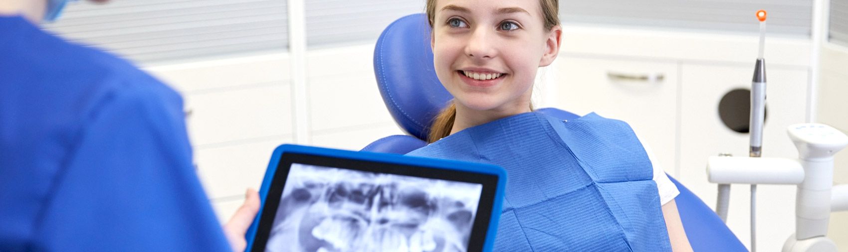
Advanced dental x rays support accurate diagnoses for effective and prompt treatment in Century City, CA
Dental issues like tooth decay, infection, and bone loss can develop beneath the gums without causing noticeable symptoms. Because these problems may not be visible during a routine visual examination, they can remain undetected until they become more serious. Digital dental X-rays give detailed images of your teeth, roots, and jawbone, which enable your dentist to detect hidden issues early. Early detection can lead to timely treatment, prevent further oral health issues, and avoid the need for more comprehensive dental treatments.
These detailed images are also used to develop a treatment plan tailored to your dental needs. A comprehensive dental exam with digital X-rays can give you valuable information about your oral health if you have tooth sensitivity, jaw discomfort, or want to maintain a healthy smile. At the dental office of Danielle Akry DDS, our general dentists use advanced digital X-rays to detect hidden dental issues early and provide personalized treatment to help maintain a healthy smile.
What Is a Dental Radiograph, and How Does It Work?
Dental radiographs provide an extremely detailed diagnostic image of your teeth, jawbone, and other surrounding structures that are completely invisible to the naked eye. A non-invasive diagnostic process that employs a very small amount of electromagnetic energy to produce detailed black, white, and gray images for a comprehensive oral health assessment.
Understanding the Science Behind X-Ray Technology
Dental X-rays use a small amount of radiation to create detailed images of your teeth, gums, and jaw. The X-ray beam passes through the soft tissues in your mouth and reaches a digital sensor. Soft tissues allow most X-rays to pass through, while denser areas, such as teeth and the jawbone, absorb more radiation.
The sensor detects these differences and sends the information to a computer, which converts it into a detailed image of your mouth. This allows dentists to see areas that may not be visible during a regular visual examination and identify potential dental problems early.
Interpretation of Radiographic Contrasts
Digital X-rays show different areas of the mouth in shades ranging from white to black. These differences help dentists identify the density of teeth, bone, soft tissues, and other structures, making it easier to detect dental issues.
- White or Light Areas (Radiopaque): Dense structures and materials that absorb most of the X-ray beam, such as enamel, dense cortical bone, metal fillings, and crowns.
- Gray Areas: Less-dense structures, including soft tissues and some tooth structures.
- Dark Areas (Radiolucent): Areas containing air or less-dense material, such as the pulp chamber, spaces within the jaw, and some areas of infection.
Understanding the Difference Between Digital Film and Traditional Film Radiography
The transition from traditional film to digital sensors has significantly improved dental imaging. Traditional X-rays required film packets and chemical processing in a darkroom, which made the process slower and less convenient. Digital X-rays eliminate the need for chemical processing by using electronic sensors connected directly to a computer, allowing images to be captured and viewed quickly at the dental chair.
These digital sensors have rounded, smooth edges. After the sensor picks up the energy beam, your image appears on a high-definition monitor. This instant visualization enables your provider to enlarge, alter the contrast, and examine each tooth as it is. Digital imaging systems use as much as 90% less radiation than film imaging systems. This significant cut makes the diagnostic process extremely safe and eliminates the toxic waste generated by the old film processing system.
Why Experienced Dentists Recommend Dental X-Rays
A visual examination can only reveal what is visible on the surface of your mouth. Modern dental X-rays allow experienced dentists to examine areas beneath the surface and detect problems such as early decay, deep infections, and structural damage before they cause severe pain or permanent tooth loss.
Finding Cavities Between Teeth
The areas where your teeth touch are common places for cavities to develop because they are difficult to see and clean. A routine visual exam may not detect decay in these narrow spaces.
Bacteria can build up between the teeth and produce acids that gradually weaken the enamel. By the time a cavity becomes visible during a regular checkup, it may have already reached the deeper dentin. Dental X-rays can reveal these cavities as small, dark areas between the teeth. Detecting decay early allows dentists to treat it with smaller fillings and preserve more of your natural tooth structure.
Detecting Decay Underneath Existing Fillings or Restorations
Structural wear occurs over years of chewing, even in the well-placed dental fillings, crowns, and bridges. Micro-gaps, which are very small, may form at the margins of these restorations, and bacteria can seep under the protective materials.
This recurrent decay is not visible on a visual exam because the filling is made entirely of metallic or ceramic material. Radiographs can pass through these physical barriers and reveal the actual condition of the tooth structure beneath older restorations. If you have an existing crown, secondary decay can be detected before it progresses into the sensitive pulp chamber of your tooth. With regular radiographic monitoring, aging restorations can be identified and replaced before they lead to severe structural damage or tooth fractures.
Checking for Bone Loss and Gum Disease
Periodontal disease is a bacterial infection that can slowly damage the gums and the bone supporting your teeth. Because this bone is hidden beneath the gums, dentists use a periodontal probe to measure the depth of gum pockets. These measurements are combined with X-rays that show bone levels to help diagnose and determine the stage and severity of periodontitis, in line with guidelines from the American Academy of Periodontology (AAP).
A dental X-ray offers a precise physical measurement of your bone levels and will give you the exact height and density of your jawbone. This imaging will show if there is horizontal bone loss along your dental arch or vertical bone loss in deep pockets.
Dental X-rays can be used to monitor bone levels, allowing dentists to catch bone loss early and tailor periodontal treatment to prevent teeth from becoming loose. These radiographic images are also helpful in assessing the effectiveness of your bone grafting procedure and the strength and stability of your jaw.
Identifying Root Infections, Cysts, and Abscesses
A tooth infection can develop if bacteria invade the pulp, infecting the nerves and blood vessels within the tooth. The infection can spread up the root canal and into the surrounding bone, causing a painful abscess to develop.
These infections occur beneath the surface and cannot be detected in a visual examination of the teeth. These infections are seen as dark areas around the tooth root on dental X-rays. Early detection allows your dentist to treat the infection with a root canal and prevent a cyst from affecting nearby teeth.
Tracking Jaw Development and Impacted Teeth
Dental X-rays are a valuable tool for tracking the growth and development of teeth and jaws from childhood to adulthood. They can demonstrate the development of permanent teeth under the baby teeth and if they are growing in the correct position.
These images can also be used to determine whether any teeth are impacted, such as wisdom teeth that are still stuck under the gums or jawbone, because there is not enough room. X-rays show where the impacted teeth are, how they relate to nearby nerves, and how they affect other teeth. This information is useful for dentists and orthodontists to plan safe extractions and orthodontic treatment.
Clinical Classifications of Dental X-Rays
There are two types of dental X-rays, depending on the sensor’s location. Intraoral X-rays are taken within the mouth and provide detailed views of each tooth; extraoral X-rays produce wider images of the jaws and surrounding structures. Both are used to diagnose issues and to determine treatment plans.
Intraoral Imaging Techniques
Intraoral X-rays use a digital sensor placed inside the mouth to capture detailed images of the teeth, roots, and supporting tissues. They are frequently used at regular dental visits.
- Bitewing Radiographs
Bitewing X-rays show both your upper and lower back teeth in a single image. You simply bite down on a small sensor to reveal cavities between teeth and evaluate supporting bone levels. These X-rays focus on the premolars and molars, where cavities commonly develop. Bitewing X-rays are most commonly performed every 12 to 18 months as part of a patient’s routine preventive care.
- Periapical Radiographs
The periapical X-rays show the entire tooth from the crown to the root and the surrounding bone. These are frequently applied when a patient has tooth pain, swelling, or deep decay. These images are useful for diagnosing root infections, fractures, and bone damage, as well as assessing the outcome of root canal therapy. They also enable dentists to track healing following dental treatments.
- Occlusal Radiographs
Occlusal X-rays use a larger sensor to capture a single image of the entire upper or lower dental arch. They help dentists examine the palate, the floor of the mouth, and the position of developing or impacted teeth. These X-rays can also detect extra teeth, cleft palate, salivary gland stones, jaw fractures, and injuries to the front teeth. Because they cover a larger area, occlusal X-rays can provide information that smaller dental X-rays may miss.
Extraoral Imaging Techniques
Extraoral X-rays are taken outside the mouth and provide a wider view of the jaws, skull, and facial bones. They are especially useful for planning orthodontic treatment, preparing for oral surgery, and evaluating overall jaw health.
- Panoramic Radiographs
A machine rotates around your head to produce a panoramic X-ray that shows the whole mouth. They display all the teeth, the upper and lower jaws, the sinuses, and the jaw joints. While they cannot detect small cavities, panoramic X-rays can help identify impacted wisdom teeth, jaw fractures, cysts, sinus issues, and other structural problems. These radiographs are often prescribed before orthodontic treatment, oral surgery, and new patient exams.
- Cephalometric Projections for Side-Profile Jaw Planning
Cephalometric X-rays are taken from the side of the head, jaws, teeth, and face. These images help orthodontists determine the relationship between the jaws and the skull. They are especially useful for planning braces, correcting bite problems such as overbites and underbites, and monitoring jaw development in children and teens. These detailed pictures help ensure that treatment is carefully planned for proper function and long-term results.
- Cone Beam CT (CBCT) Scans for 3D Oral Imaging
Cone Beam Computed Tomography (CBCT) provides advanced three-dimensional dental imaging technology. The CBCT scanner rotates around your head, taking hundreds of high-resolution images in just a few seconds. These images are then combined into a highly detailed 3D model of the entire teeth, jaw, and facial structures by an advanced computer program.
This 3D model enables your dentist to see the bone structures, teeth, and crucial nerve pathways from any perspective, offering much more spatial context than traditional flat images. CBCT scans are essential for planning dental implant placement because they enable dentists to accurately measure bone volume. They also help to map out complex root canals, evaluate TMJ disorders, and plan safe and predictable oral surgeries.
Prioritizing Safety and Modern Radiation Guidelines
In modern dental practices, patient safety is the number one rule. Dentists use the latest technology and adhere to the latest clinical protocols to minimize your radiation exposure.
Low-Dose Radiation and Digital Sensor Standards
The safety principle known as “As Low As Reasonably Achievable” (ALARA) is used to minimize radiation exposure during dental X-rays. This means taking every reasonable precaution to use the lowest radiation dose needed to produce clear, useful images. This approach is consistent with recommendations for radiation safety in dental care.
Digital X-ray sensors can significantly reduce radiation exposure compared with traditional film X-rays. Because digital sensors are highly sensitive, they can capture clear images quickly with less radiation. The equipment can also be adjusted based on factors such as the patient’s age, size, and the area being examined. These measures help limit radiation exposure while providing the detailed images needed for an accurate diagnosis.
Updates on Lead Aprons and Thyroid Collars, 2024–2026
Current radiation safety guidance focuses on minimizing radiation exposure by using proper equipment, beam collimation, and the lowest exposure settings needed to produce a clear diagnostic image. The FDA states that lead aprons are not required for routine dental X-rays when other radiation-reduction measures are followed. A thyroid shield may be used when it does not interfere with the diagnostic image and is particularly essential for children.
Modern digital X-ray systems use sensitive image receptors and tightly controlled X-ray beams to limit unnecessary radiation exposure. Proper collimation keeps the X-ray beam focused on the area being examined. The right exposure settings help produce clear images while using the lowest practical radiation dose. The FDA also recommends following the ALARA principle and using beam collimation, appropriate exposure settings, and the minimum number of images needed for diagnosis.
Protective shields can sometimes interfere with the X-ray beam and degrade image quality if not positioned correctly. A thyroid collar may be used when the thyroid gland could be exposed, but it should not block or interfere with the area being examined. These measures help reduce unnecessary radiation exposure while allowing dentists to obtain clear images required for accurate diagnosis and treatment planning.
Safe Dental Imaging During Pregnancy
Excellent oral health is crucial during pregnancy because active gum infections and tooth decay can affect the health of both mother and baby. Diagnostic dental radiographs are safe and necessary during pregnancy when used with standard clinical precautions and do not pose any measurable risk to the developing fetus.
The X-ray beams used in modern dentistry are very small and do not directly affect your abdomen or reproductive organs. Untreated dental infections can cause significant pain, systemic inflammation, and severe medical complications.
Your pregnancy status should be discussed with your dental team before taking X-rays. When dental imaging is necessary, appropriate safety measures can help protect you and your developing baby while allowing your dentist to make an accurate diagnosis.
What To Expect During The Procedure
Dental X-rays are quick, painless, and very organized. Your dentist should strive to make you feel relaxed and comfortable during the short imaging procedure. There are almost no special preparations or dietary restrictions for dental X-rays.
Please brush your teeth thoroughly before going to the clinic to help create a clean, hygienic environment for your clinical team. California Code of Regulations (CCR) Title 17, Section 30100, requires dental practices to have structural and clinical safety measures in place to ensure all radiographic procedures are performed under the direct supervision of licensed personnel.
Upon arrival, your dental provider will ask you to remove any metal accessories from your head and neck. These include things like dangling earrings, facial piercings, bobby pins, metal-framed glasses, or removable orthodontic retainers. Metal items completely absorb radiographic energy and appear as bright white obstructions that may obscure underlying decay or anatomical structures.
Step-by-Step Walkthrough of the Imaging Session
Getting your dental X-rays is quick and painless, taking only a few minutes. Your dentist will guide you through each stage of your appointment with a caring and patient approach.
- Comfortable Seating: You sit in a comfortable dental chair while an assistant adjusts the chair to fit you.
- Metal Object Removal: Your dental provider will double-check for metal objects on the face or in glasses to avoid overall distortion in the radiograph.
- Sensor Placement: Your dentist will carefully insert a soft digital sensor directly into your mouth.
- Stay Still: Your dentist could ask you to bite down lightly as they activate the machine from behind the protective wall shield.
What Happens After Your X-Rays Are Taken
When the imaging machine is activated, the electronic signal is sent directly to a computer beside the dental chair. The high-resolution digital X-ray appears on the screen within seconds. Unlike traditional film X-rays, digital images do not require chemical processing, which saves time and helps keep appointments efficient.
After the images are taken, your general dentist will review them with you and explain what they show. Digital X-rays can help identify healthy bone levels, detect early signs of decay, and determine whether treatment is needed. Reviewing the images together helps you understand your oral health and stay involved in decisions about your dental care.
Frequently Asked Questions about Oral Radiographs
Questions about the safety, clinical need, and diagnostic value of dental X-rays are completely understandable. Your dentists can help you make informed decisions regarding your oral care.
Can a Patient Legally Refuse Dental X-Rays?
Yes, as a patient, you have the legal right to refuse any diagnostic procedure, including dental radiographs. You should remember, however, that refusing to undertake necessary X-rays prevents your dentist from delivering the full standard of dental care. A dentist may not be able to detect hidden bone infections, cavities between teeth, or problems affecting the tooth roots without diagnostic images.
Dental X-rays can provide essential information needed to plan treatments such as fillings, extractions, and deep cleanings. Without the appropriate imaging, it may be difficult to diagnose certain conditions accurately or determine the safest treatment. If you have concerns about radiation exposure, your dentist can explain why specific X-rays are recommended and how they help guide your care.
Can Dental X-rays Detect Oral Cancers and Tumors?
Dental radiographs are very useful diagnostic tools and may show advanced pathological conditions such as cysts, non-cancerous tumors, and cancers of the jawbone. These lesions appear as abnormal dark (radiolucent) or light (radiopaque) areas in the jawbone on a panoramic radiograph or CBCT scan.
Traditional dental X-rays, however, will not detect cancer in your tongue, throat, cheeks, or floor of your mouth. A thorough visual and physical examination of the soft tissues of the mouth during a routine dental checkup is necessary to diagnose soft-tissue oral cancer.
At your appointment, your dentist will thoroughly examine all of your soft tissues for any unusual white, red, or sore areas, lumps, or patches. A thorough physical examination, together with state-of-the-art digital X-rays, provides a powerful, highly protective measure against oral disease.
Consult an Experienced General Dentist Near Me
Routine checkups and digital X-rays are essential for maintaining good oral health, as they can help identify potential issues before they worsen. Early diagnosis means early treatment, which helps to maintain your teeth and avoid more complicated dental treatment. A dentist who uses advanced technology and provides personalized care can make a significant difference in your oral health.
At the Danielle Akry DDS dental clinic, we are dedicated to providing you and your family in Los Angeles with comprehensive dental care that meets your unique needs. Contact us today at (310) 286-3111 to schedule your dental checkup or discuss any concerns about your oral health.